
Fig. S1
- ID
- ZDB-FIG-181116-32
- Publication
- Koch et al., 2018 - Intestinal microbiome adjusts the innate immune setpoint during colonization through negative regulation of MyD88
- Other Figures
- All Figure Page
- Back to All Figure Page
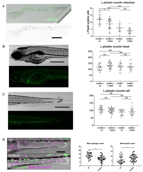
(A) Excised larval intestines conrm statistically elevated numbers of L-plastin positive cells in germ-free intestines. No signicant dierence was observed in the intestines of Myd88 decient larvae. (mean � s.e.m, n=2 biological replicates, 6-8 embryos per group), *p0,05; ***p0,001 by two-way ANOVA with Bonferroni correction for multiple comparisons. (B-C) Whole embryo assessment of overall leukocyte assessment by L-plastin immunostaining, (mean � s.e.m, n=2 biological replicates, 6-8 embryos per group) Acquired by confocal microscopy at 5x magnication, no signicant dierences were observed by two-way ANOVA with Bonferroni correction for multiple comparisons. (D) Quantication of macrophages (magenta) and neutrophils (green) in the distal intestine of GF versus CONVD wt larvae of the Tg(Mpx:GFP/Mpeg:mCherryF) line)at 5 DPF, acquired by confocal microscopy at 20x magnication. The region of analysis is outlined in white lines, (mean � s.e.m, n=3 biological replicates, 6-8 embryos per group) *p0,05; ***p0,001 by Student?s t-test. Scalebars, 200 ?m in A; 500 ?m in B-C; 100 ?m in D. |
| Fish: | |
|---|---|
| Condition: | |
| Observed In: | |
| Stage: | Day 5 |