
Fig. 5
- ID
- ZDB-FIG-180911-36
- Publication
- Muralidharan et al., 2018 - Retinal Wnt signaling defect in a zebrafish fetal alcohol spectrum disorder model
- Other Figures
- All Figure Page
- Back to All Figure Page
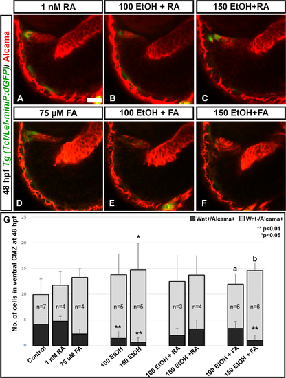
RA and FA co-supplementation rescue Wnt signaling. Alcama staining on Tg(Tcf/lef-miniP:dGFP) fish showed that ethanol treatment reduced the number of Wnt+/Alcama+ cells in the peripheral CMZ. Control and ethanol treated embryos in this experiment appeared identical to those shown in Fig 4A–4C and were left out for brevity. (A-F) RA co-supplementation could rescue Wnt+/Alcama+ cells (A-C); FA co-supplementation could also rescue Wnt+/Alcama+ cells in the CMZ particularly in 100 mM Ethanol + FA treated fish (D-F). (G) Quantification of Wnt+/Alcama+ cells in the peripheral CMZ showed significant reduction after ethanol treatment, which was rescued by RA and FA co-supplementation. Numbers of cells were counted in a single optic-nerve containing confocal optical section in the ventral CMZ. Images show rostral at top, lateral at left. Error bars indicate standard deviation. ‘**’ indicates statistical significance in comparison to control embryos (p<0.01). ‘*’ indicates statistical significance in comparison to control embryos (p<0.05). ‘a’ indicates statistical significance in comparison to 100 mM ethanol embryos (p<0.05). ‘b’ indicates statistical significance in comparison to 150 mM ethanol embryos (p<0.05). Scale bar = 10 μm for panels A-F. |
| Fish: | |
|---|---|
| Conditions: | |
| Observed In: | |
| Stage: | Long-pec |