
Fig. S8
- ID
- ZDB-FIG-180827-44
- Publication
- Astone et al., 2018 - Zebrafish mutants and TEAD reporters reveal essential functions for Yap and Taz in posterior cardinal vein development
- Other Figures
- All Figure Page
- Back to All Figure Page
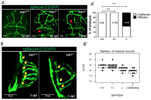
yap1-/- larvae exhibit cranial and hyaloid vasculature defects. (A) Representative figures of yap1 mutants showing cranial vasculature defects compared to WT sibling at 72 hpf. The red arrows point to lack of endothelial transgene expression in the mesencephalic vein (MsV) and dorsal longitudinal vein. (A') The cranial vasculature phenotype is partially penetrant. (B) Dorsal view of the eyes of 5 dpf WT and mutant siblings. (B') Number of hyaloid vessels per embryo for each genotype. Each point represents one eye. The number of hyaloid vessels of wild-type embryos is statistically higher in comparison to mutants with (p<0.01) or without (p<0.05) coloboma by student T test. n for each group is indicated. *** = p<0.001. |
| Fish: | |
|---|---|
| Observed In: | |
| Stage Range: | Protruding-mouth to Day 5 |