Fig. 2
- ID
- ZDB-FIG-180622-4
- Publication
- Schepis et al., 2018 - Protease signaling regulates apical cell extrusion, cell contacts, and proliferation in epithelia
- Other Figures
- All Figure Page
- Back to All Figure Page
|
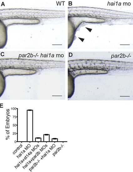
Par2b deficiency prevents grossly abnormal skin morphology in hai1a morphants. (A?D) Wild-type or par2b?/? embryos were injected with the indicated MOs and imaged at 30 hpf. Images of control (A), hai1a morphant (B), par2b?/?:hai1a morphant (C), and par2b?/? embryos (D) are shown. Note the presence of cell clusters on hai1a morphants (B, arrowheads) and their absence on controls and Par2b-deficient hai1a morphants. Bar, 200 Ám. (E) Percentage of embryos with cell clusters. 100 or more embryos were analyzed for each condition; mean ▒ SEM for three independent experiments is shown. Statistical analysis was performed using one-way ANOVA and Bonferroni posttest. The hai1a morphant was different from all other groups (P < 0.0001). |
| Fish: | |
|---|---|
| Knockdown Reagents: | |
| Observed In: | |
| Stage: | Prim-15 |

