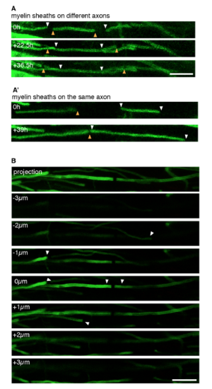
Fig. S4
|
Timelines of myelin sheath growth to identify neighbouring sheaths from confocal zstacks in the spinal cord of mbp transgenic zebrafish. Related to STAR Methods. A) Nearby myelin sheaths (arrowheads) are shown that do not oppose another over time in the x/y axis to form a nodal gap, and which must therefore locate on different axons. A?) Nearby myelin sheaths similar to A, but which do oppose another over time in the x/y axis to form a nodal gap, and which are therefore considered to locate on the same axon. Scale bar: 10 ?m B) Maximum intensity projection and single planes of a confocal z-stack of the myelin sheaths shown in Figure 6A. Arrowheads depict nearby sheath when they are in the same focal plane. |

