Fig. 6
|
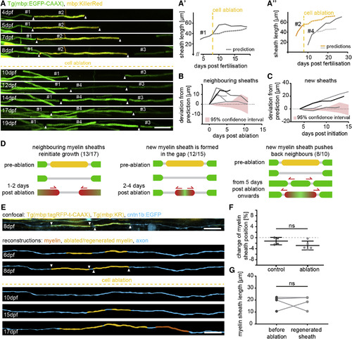
Myelin Sheaths Dynamically Remodel after Targeted Oligodendrocyte Ablation (A?A?) Timeline of confocal images of the formation of three consecutive sheaths, ablation, and subsequent remodeling between 4 and 19 days post-fertilization (dpf). (A?) shows the quantification of growth curves of sheath #1 over the time of analysis and its predicted growth (dotted line) based on its age and animal body growth. (A?) shows the quantifications of the measured sheaths #2 and #4 as seen in (A), and the respective predicted growth (dotted lines) described in (A?). Scale bar, 10 ?m. (B) Quantification of deviated growth of individual sheaths neighboring the ablation site. (C) Quantification-deviated growth of new sheaths formed in the ablation site. (D) Schematic summarizing the frequency of observed/possibly observable events of sheath growth and remodeling. (E) Top: confocal image of discontinuously myelinated axons labeled with cntn1b:EGFP in a Tg(mbp:KR), Tg(mbp:tagRFPt-CAAX) transgenic animal at 8 dpf. Below: reconstructions of the same axon shown above at different time points before and after oligodendrocyte ablation between 6 and 17 dpf. Scale bar, 10 ?m. (F) Quantification of the relative position of regenerated sheaths and the relative position of control sheaths along discontinuously myelinated axons over the same time. Data are expressed as mean ▒ SD (t test, t(7) = 1.598, p = 0.15). (G) Quantification of sheath length before and after ablation along discontinuously myelinated axons (t test, t(3) = 0.0592, p = 0.96). See also Movies S4, S5, and S6. |

