
Fig. 4
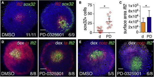
Fgf Signaling Represses Endoderm Specification around Ndr1 Clones (A) Animal views of DMSO- and PD-0325901-treated germ ring-stage embryos containing Ndr1-expressing clones stained for lft2 and sox32. (B) Quantification of sox32-positive cells in (A). Means � SD, Mann-Whitney U test; ?p < 0.05. (C) Quantification of surface area of lft2-positive domain surrounding Ndr1-expressing clones in embryos treated as in (A). n = 3, 11 embryos in total. Means � SD, two-tailed t test; ?p < 0.05. (D) Animal views of germ ring-stage embryos treated as in (A), but stained for ta and lft2. (E) Animal views of germ ring-stage embryos treated as in (A), but stained for noto and lft2. Scale bars, 100 ?m. See also Figure S4. |
Reprinted from Developmental Cell, 44(2), van Boxtel, A.L., Economou, A.D., Heliot, C., Hill, C.S., Long-Range Signaling Activation and Local Inhibition Separate the Mesoderm and Endoderm Lineages, 179-191.e5, Copyright (2017) with permission from Elsevier. Full text @ Dev. Cell