
|
Fig. 4
Fgf Signaling Represses Endoderm Specification around Ndr1 Clones
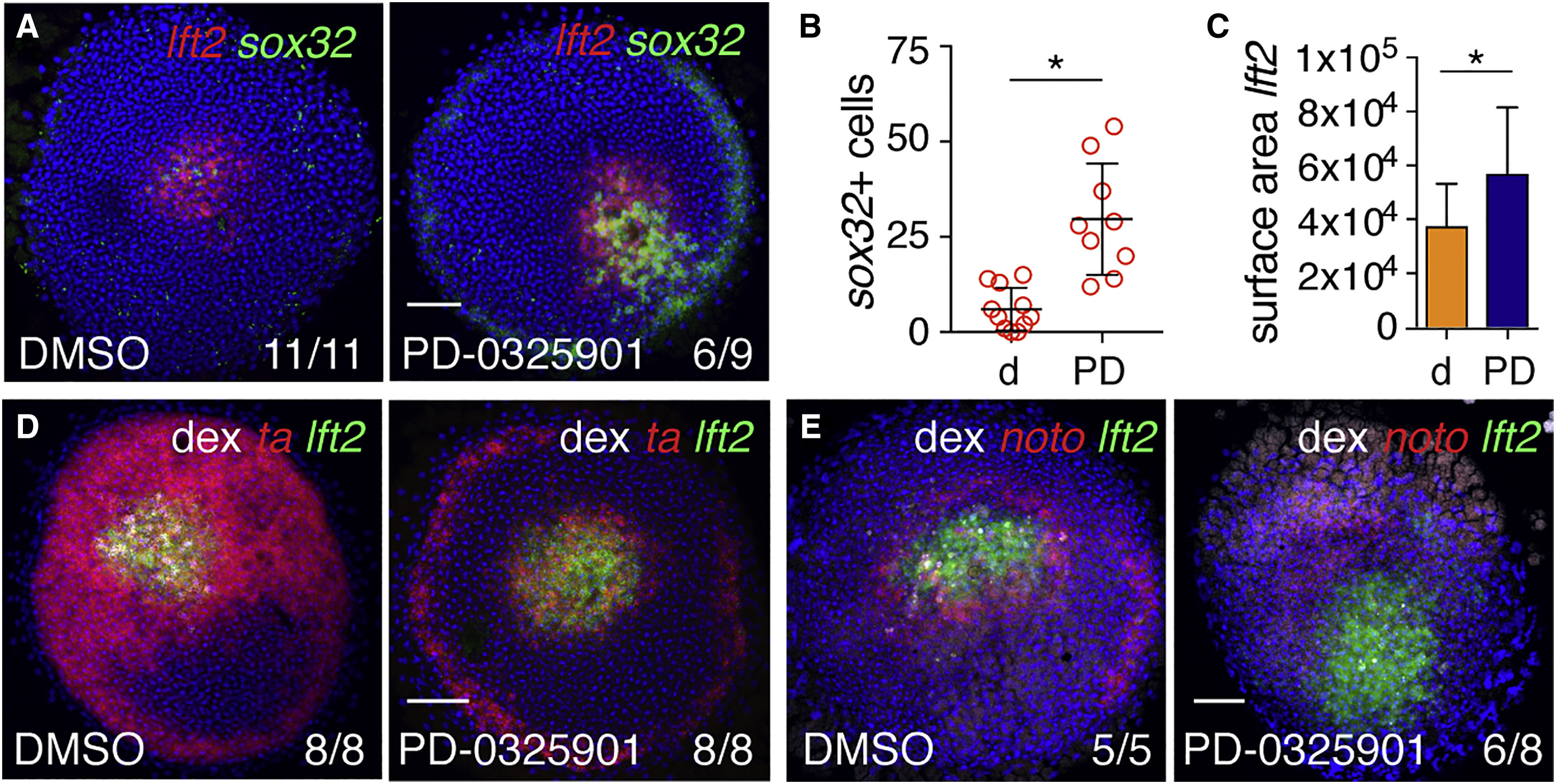
(A) Animal views of DMSO- and PD-0325901-treated germ ring-stage embryos containing Ndr1-expressing clones stained for lft2 and sox32.
(B) Quantification of sox32-positive cells in (A). Means � SD, Mann-Whitney U test; ?p < 0.05.
(C) Quantification of surface area of lft2-positive domain surrounding Ndr1-expressing clones in embryos treated as in (A). n = 3, 11 embryos in total. Means � SD, two-tailed t test; ?p < 0.05.
(D) Animal views of germ ring-stage embryos treated as in (A), but stained for ta and lft2.
(E) Animal views of germ ring-stage embryos treated as in (A), but stained for noto and lft2.
Scale bars, 100 ?m. See also Figure S4.
Reprinted from Developmental Cell, 44(2), van Boxtel, A.L., Economou, A.D., Heliot, C., Hill, C.S., Long-Range Signaling Activation and Local Inhibition Separate the Mesoderm and Endoderm Lineages, 179-191.e5, Copyright (2017) with permission from Elsevier. Full text @ Dev. Cell