
FIGURE
Fig. 1
- ID
- ZDB-FIG-180523-4
- Publication
- Fu et al., 2018 - Novel Animal Model of Crumbs-Dependent Progressive Retinal Degeneration That Targets Specific Cone Subtypes
- Other Figures
- All Figure Page
- Back to All Figure Page
Fig. 1
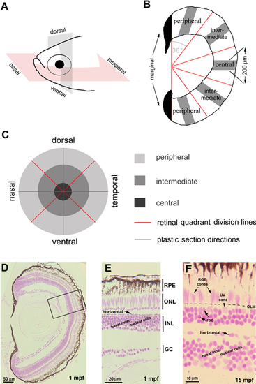
Linear cell densities of six categories of retinal cells were evaluated by JB4-Feulgen histology. (A) Zebrafish eyes were sectioned in either the nasal-temporal (anterior-posterior) or ventral-dorsal axis. (B) Each retinal section was partitioned by a vertical line to exclude the developing marginal region (black) from the differentiated retina, which was further divided into five regions each with angular subtense of 36 degrees. We counted nuclei in 200-μm linear segments (gray bars). (C) Spatial relationships among the nine sampled retinal regions. (D–F) JB4-Feulgen histology illustrates the localization and morphologies of the six retinal cell categories at 1 mpf (D, E; the inset box in D outlines the boundary of E) and 15 mpf (F). The dashed line indicates the location of the OLM. GC, ganglion cell layer; INL, inner nuclear layer; ONL, outer nuclear layer.
|
Expression Data
Expression Detail
Antibody Labeling
Phenotype Data
Phenotype Detail
Acknowledgments
This image is the copyrighted work of the attributed author or publisher, and
ZFIN has permission only to display this image to its users.
Additional permissions should be obtained from the applicable author or publisher of the image.
Full text @ Invest. Ophthalmol. Vis. Sci.