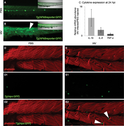
Molecular and cellular markers of inflammation are present in the muscle tissue of IAV-infected zebrafish All embryo images are side mounts, dorsal top, anterior left of zebrafish at 24 hpi. (A) PBS-injected Tg(NFkB:GFP) zebrafish embryo which expresses GFP in cells where NFkB transcription factor-dependent gene expression is occurring. NFkB signaling is active in the lateral line system, but not in muscle cells. Inset panel is a merge of fluorescence and brightfield imaging. (B) IAV-injected Tg(NFkB:GFP) zebrafish embryo. NFkB-dependent gene transcription is turned on in muscle cells in response to IAV infection. Inset panel is a merge of fluorescence and brightfield images. (C) Quantification of pro-inflammatory cytokine mRNA expression in IAV-infected zebrafish compared to PBS-injected zebrafish at 24 hpi. Expression of interleukin 1, beta (11.1 +/- 5.2-fold increase; 3 biological replicates; 3 independent experiments) and interleukin 8 (3.7 +/- 1.4-fold increase; 3 biological replicates; 3 independent experiments) increases in response to IAV while tumor necrosis factor a expression remains unchanged (1.1 +/- 0.5-fold increase; 2 biological replicates; 2 independent experiments). (D-E2) Lettered panels show phalloidin staining (red), panels numbered 1 show GFP-positive neutrophils, and panels numbered 2 are merged. (D-D2) PBS-injected Tg(mpx:GFP) zebrafish. Note that not many neutrophils are present in muscle tissue. (E-E2) IAV-infected Tg(mpx:GFP) zebrafish. Note the retracted muscle fibers and the infiltration of muscle tissue by neutrophils. White arrowheads in E2 point to neutrophils localized to the unanchored ends of retracted muscle fibers.
|

