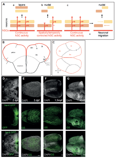
Fig. s1
- ID
- ZDB-FIG-180403-41
- Publication
- Furlan et al., 2017 - Life-Long Neurogenic Activity of Individual Neural Stem Cells and Continuous Growth Establish an Outside-In Architecture in the Teleost Pallium
- Other Figures
- All Figure Page
- Back to All Figure Page
|
Eversion exposes the germinative RG layer in the adult zebrafish pallium, related to Figure 1. A. Schematic representations of the neural stem cell (NSC)-dependent (a, b) and -independent (c) strategies shaping brain cytoarchitecture. The germinal NSC pool and its neurogenic activity are indicated in red, and neuronal identities issued from this pool are colorcoded. The output structure is depicted at the top of each panel. (a) Continuous NSC activity generates age-related layers in the mammalian neocortex, in an inside-out fashion following radial neuronal migration. (b, c) Spatially controlled NSC activity and neuronal migration events organize functional nuclei in the bird and reptile neocortex (from [S4][S5]) B. Simplified schematized cross section of the zebrafish adult pallium at medial antero-posterior levels indicating the parenchymal subdivisions and sulci referred to in this study (from [S1]). The attachment point of the tela choroida, positioning the limit of the eversion process, is indicated (gray dots and arrows) (from [S2]). The pallial germinative zone (ventricular zone, red) extends from this point to the pallial-subpallial boundary (gray dashed line). C. Simplified schematized whole-mount view of the zebrafish adult pallium (viewed from top, anterior left) indicating the germinal zone areas visible from a dorsal view, as identified in [S3]. D-G. Whole-mount dorsal views of the pallial germinal zone in transgenic her4:eGFP telencephali, identified by the expression of GFP or GS (as indicated) at 2dpf (D), 5dpf (E), 1.5mpf (F) and 3mpf (adult, G). Whole-mount preparations counterstained with DAPI. Abbreviations: Da: anterior pallium; Dc: central pallium; Dl: lateral pallium domain; Dm: medial pallium domain; Dp: posterior pallium domain; ls: lateral sulcus; t.c.: tela choroida; sy: sulcus ypsiloniformis. Scale bars: D, E : 20?m; F : 50?m; G : 100?m. |

