
Fig. 2
- ID
- ZDB-FIG-180403-35
- Publication
- Furlan et al., 2017 - Life-Long Neurogenic Activity of Individual Neural Stem Cells and Continuous Growth Establish an Outside-In Architecture in the Teleost Pallium
- Other Figures
- All Figure Page
- Back to All Figure Page
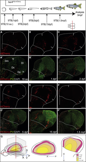
Zebrafish Pallial Neurogenesis Follows a Sequential Stacking Process: Medio-lateral Analysis Top: experimental design. (A?F?) Distribution of mCherry-positive neurons (A?F) born from her4-positive RGs in her4H2a-mCherry induced with 9TB at the stages indicated. Cross-sections at 3 mpf at mid-antero-posterior levels, the level indicated by a red line on telencephalon dorsal view, are co-labeled for parvalbumin (PV; A??F?). Solid white lines indicate pallium-subpallium boundary. Scale bar, 50 ?m. (G) Color-coded map of the position in the adult brain (left: ?open? whole-mount view; middle: horizontal section; right: cross-section) of the neurons born from her4-positive RGs at the stage indicated. See also Figure S4. |