Fig. 6
- ID
- ZDB-FIG-180321-7
- Publication
- Hozumi et al., 2017 - Nuclear movement regulated by non-Smad Nodal signaling via JNK is associated with Smad signaling during zebrafish endoderm specification
- Other Figures
- All Figure Page
- Back to All Figure Page
|
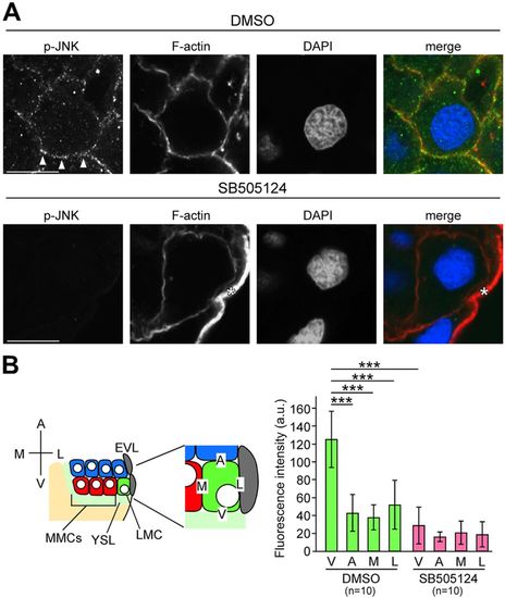
JNK is activated by Nodal from the YSL. (A) Transverse sections of DMSO-treated control and SB505124-treated embryos; p-JNK and F-actin were visualized by immunohistochemical staining and phalloidin, respectively at 4.7 hpf. Fluorescent signals are displayed as grayscale images. Arrowheads indicate p-JNK fluorescence on the boundary between the YSL and LMCs. Asterisk indicates the cell membrane of EVL cells. Scale bars: 10 µm. (B) Fluorescence intensities were measured along the cell membrane on the boundaries between the YSL and LMCs (V), on cells located animal-pole side and LMCs (A), EVL and LMCs (L), or MMCs and LMCs (M). Error bars represent s.d. of ten measurements. n, number of nuclei examined. ***P<0.001. |

