FIGURE

Fig. S5

ID
ZDB-FIG-180221-34
Publication
Wakisaka et al., 2017 - An Adenosine Receptor for Olfaction in Fish
Other Figures
All Figure Page
Back to All Figure Page
Fig. S5

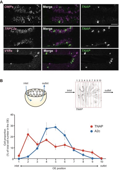
TNAP is expressed in non-neuronal cells predominantly located in the anterior OE close to the inlet naris. Related to Figure 6.
(A) Double fluorescence in situ hybridization with cRNA probes for TNAP (green) and OMPs, TRPC2s, or V1Rs (magenta) on OE sections. No overlap is observed for TNAP signals with any OSN markers, indicating that TNAP is expressed in non-neuronal cells in the OE (n=3). Scale bars, 20 μm.
(B) Distribution of TNAP- and A2c-expressing cells in the zebrafish OE. Top left, a schematic diagram showing water flow through the zebrafish OE. Top right, a representative in situ hybridization image of TNAP expression in an OE section. Each OE section was divided into 10 compartments along the longitudinal axis and the numbers of TNAP and A2c-positive cells were counted. Bottom, a graph showing distribution of TNAP (red)- and A2c (blue)-expressing cells in the OE (n=3). Note that TNAP-expressing cells are predominantly located in the vicinity of the OE inlet, whereas A2c-expressing OSNs are observed in the middle portion between the inlet and outlet.

Expression Data

Expression Detail
Antibody Labeling
Phenotype Data

Phenotype Detail
Acknowledgments
This image is the copyrighted work of the attributed author or publisher, and ZFIN has permission only to display this image to its users. Additional permissions should be obtained from the applicable author or publisher of the image. Full text @ Curr. Biol.