Fig. S5
|
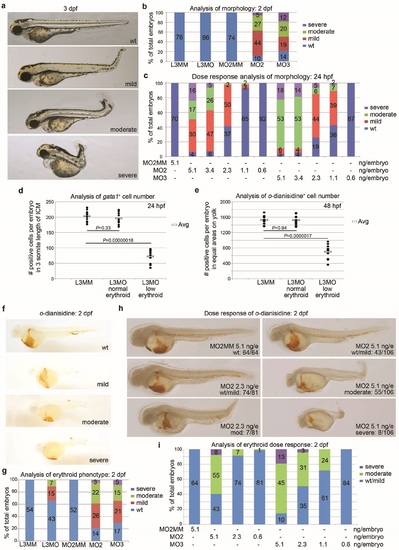
Phenotype analysis of drl family morphants. (a) Brightfield microscopy of embryos at 3 dpf with morphology defined as wild type (wt), mild, moderate, and severe as listed. Representative embryos are control (wt) or M02-injected morphants. (b) Percent of morpholino-injected embryos at 2 dpf that display developmental defects. Phenotypes defined in (a). Control mismatch morpholinos = L3MM and M02MM. The number of embryos is shown in the columns. (c) Embryos injected at the indicated dose were scored for severe, moderate, mild or normal body axis length/amount of tissue as outlined in panel a. Percent of total embryos is charted, with the embryo number shown in the column. (d) Quantitation of 24 hpf gata1-expressing cells in a 3-somite span of ICM (somites 10-13, counting from anterior) in morphants appearing to have normal (L3MM, L3MO normal) or decreased (L3MO decreased) cell numbers as labeled. (e) Quantitation of 48 hpf o-dianisidine stained cells in an equal area and region on the (left side) yolk in morphants appearing to have normal (L3MM, L3MO normal) or decreased (L3MO decreased) cell numbers as labeled. All embryos scored displayed blood pooling on the left side of the yolk. Scatter charts show cell numbers in individual embryos (closed circles); 1 O embryos per group. Average cell number per embryo is indicated (open box). Significance was determined by 2-tailed paired Student's t-test. (f) 0-dianisidine staining of control (wt) and zebrafish M02 morphants. (g) The quantitation of embryos showing wild type, mild, moderate or severe decreases in mature erythroid cells. Phenotypes defined in (f). (h) Representative images of 48 hpf o-dianisidine stained embryos injected with the indicated doses of M02MM and M02. Phenotype scoring is indicated: Wild-type=wt; moderate and severe. Note that the severity of the morphological defects correlate with the severity of the erythroid defect. (i) Quantitation of the embryos with the o-dianisidine phenotypes outlined in (h) injected with the indicated doses of morpholino. |

