
Fig. 3
- ID
- ZDB-FIG-171206-85
- Publication
- Wan et al., 2017 - Characterization of ?? T Cells from Zebrafish Provides Insights into Their Important Role in Adaptive Humoral Immunity
- Other Figures
- All Figure Page
- Back to All Figure Page
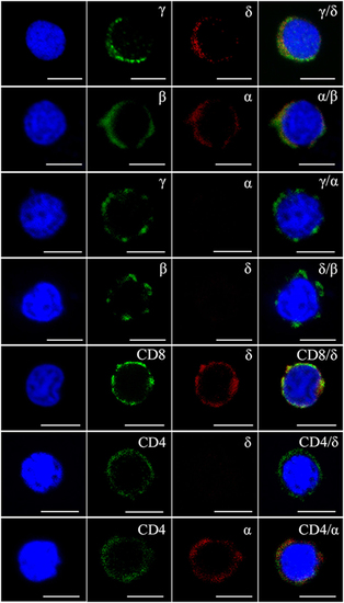
Figure 3. Immunophenotype analysis of zebrafish ?? T cells by Abs against various surface molecules. The magnetic sorted cells were double stained with anti-?, anti-?, anti-?, anti-?, anti-CD8, and anti-CD4 Abs in different combinations (rabbit anti-?/mouse anti-?, rabbit anti-?/mouse anti-?, rabbit anti-?/mouse anti-?, rabbit anti-?/mouse anti-?, rabbit anti-CD8/mouse anti-?, rabbit anti-CD4/mouse anti-?, and rabbit anti-CD4/mouse anti-?, respectively). Positive control cells were stained with rabbit anti-? and mouse anti-? Abs. Non-related Abs, including mouse IgG and rabbit IgG, were used as negative controls (data not shown). DAPI stain showed the locations of the nuclei. A laser-scanning confocal microscopy (Zeiss LSM-710) was used in the analyses (original magnification � 630, scale bar, 5 �m). |