
Fig. S9
- ID
- ZDB-FIG-171127-31
- Publication
- Ota et al., 2016 - Functional visualization and disruption of targeted genes using CRISPR/Cas9-mediated eGFP reporter integration in zebrafish
- Other Figures
- All Figure Page
- Back to All Figure Page
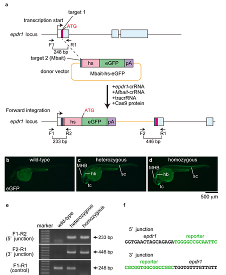
The eGFP expression in Tg[epdr1-hs:eGFP] embryos. (a) A schematic representation of the epdr1 locus containing the 5' untranslated region (white box) and the epdr1-crRNA target site (pink box), and the reporter construct consisting of the Mbait-crRNA target site (blue box), the hsp70 promoter (hs, light red box), the eGFP gene (green box) and the polyA signal (pA; purple box). (b-d) eGFP expression in wild-type, heterozygous and homozygous Tg[epdr1-hs:eGFP] embryos. (e) The integration of the reporter into the epdr1 locus was determined by genomic PCR using the epdr1-specific and the reporter-specific primers. (f) Sequence of the 5' junction and the 3' junction at the integration site of Tg[epdr1-hs:eGFP]. Black letters and green letters represent epdr1 sequences and reporter sequences, respectively. |