
Fig. 4
- ID
- ZDB-FIG-171113-32
- Publication
- Powell et al., 2016 - Zebrafish M�ller glia-derived progenitors are multipotent, exhibit proliferative biases and regenerate excess neurons
- Other Figures
- All Figure Page
- Back to All Figure Page
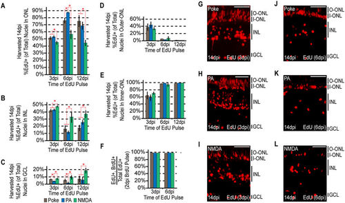
The ultimate localization of MG-derived progenitors becomes progressively more biased at later rounds of proliferation. Fish were given an intraperitoneal injection of BrdU at 2 dpi, followed by an injection of EdU at 3, 6, or 12 dpi. Each sample was harvested at 14dpi. (A?C) EdU+ nuclei were counted at the times indicated and the percentage of EdU+ nuclei residing in the (A) ONL, (B) INL and (C) GCL was determined for each injury model. Data represents means?�?s.d. (n???3). *P?<?0.02941. (D,E) The percentage of ONL EdU+ nuclei residing in (D) the upper region or (E) the lower region was determined for each injury model. (F) Co-staining samples for BrdU and EdU demonstrates that the cells proliferating at later times are a subpopulation of those proliferating at earlier times. (G?L) Representative images of retinal sections analyzed in (A?F) that were stained for EdU at (G?I) 3 dpi or (J?L) 6 dpi following (G,J) needle poke, (H,K) PA or (I?L) NMDA injury. Scale bar is equal to 50??m. Abbreviations are as in Fig. 1. |