
Fig. S5
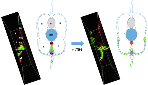
Scheme of the migration path of neutrophils from the CHT induced by bath addition of LTB4. While embryos were imaged in lateral view as in Fig. 5D,E and Movie 6, this scheme depicts the same caudal region but in cross section, so as to show the migration path of neutrophils (green) initially in the CHT, between caudal artery (ca) and vein (cv), upon bath addition of LTB4; they migrate beneath the skin that overlies the ventral fin (vf) and somitic muscles (s). To the left of each drawing is a similarly oriented view of neutrophil distribution in a live wt embryo before and after LTB4, obtained by orthogonal projection of the confocal image stacks acquired in lateral view from the left side, as shown in maximal projection in Fig. 5D and Movie 6 (left panel). Ntc, notochord; sc, spinal cord. |