
Fig. S2
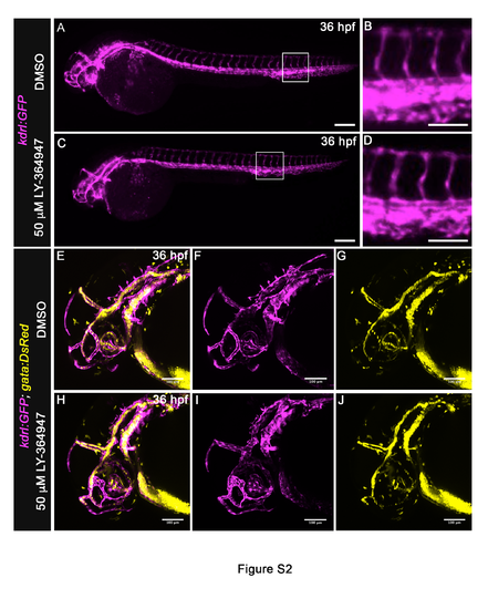
Inhibition of TGF? signaling does not grossly disrupt the body or cranial vasculature, Related to Figure 1. (A-D) Lateral views of 36 hpf Tg(kdrl:GFP) embryos treated from 20 hpf with DMSO (A,B) or 50?M LY-364947 (C,D) imaged in the green channel on a compound microscope to visualize endothelium (pseudocolored magenta). The body vasculature appeared similar between LY-364947-treated and DMSOtreated embryos. Magnified views (B,D) of boxed regions (A,C) highlight intersomitic vessels. (E-J) Left lateral views of 36 hpf Tg(kdrl:GFP), Tg(gata1:DsRed) embryos treated from 20 hpf with DMSO (E-G) or 50?M LY-364947 (H-J) imaged on a confocal microscrope in the green and red channels to visualize endothelium (pseudocolored magenta) and red blood cells (RBC) (pseudocolored yellow), respectively. Merged (E,H) and single channel (F,G,I,J) images are shown. In both cohorts, cranial vascular development and integrity appeared grossly normal and supported RBC circulation. (A,C) Scale bars = 200?m, (B,D-J) Scale bars = 100?m. (n=4 to 5 embryos per group) |
| Genes: | |
|---|---|
| Fish: | |
| Condition: | |
| Anatomical Terms: | |
| Stage: | Prim-25 |