
Fig. 3
- ID
- ZDB-FIG-170825-20
- Publication
- Orr et al., 2016 - A mutation in the atrial-specific myosin light chain gene (MYL4) causes familial atrial fibrillation
- Other Figures
- All Figure Page
- Back to All Figure Page
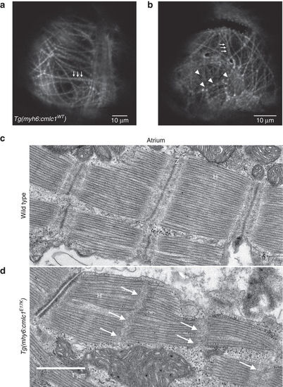
Myofibrillar organization and sarcomeric structure in WT and E17K transgenics. (a) A WT transgenic atrium at larval stage (5 d.p.f.) in the Tg(myl7:LifeAct-GFP)s974 background shows GFP-labelled F-actin, with myofibrils organized linearly across the atrium, either up-and-down or left-to-right. The myofibrils have a ?dotted line? pattern, with well-demarcated box-like shapes indicating normal arrangement of F-actin in the myofibrils. The non-fluorescent gaps (arrows) represent the actin-poor H-zones. (b) In an E17K transgenic larval atrium, some myofibrils appear unaffected (arrows), but there are large areas where myofibrils are not linearly organized and actin localization appears abnormal, giving a stippled appearance to the sarcomeres without clear H-zones (arrowheads). (c) Electron microscopy of adult WT zebrafish atrium shows clear H-zones and Z-disks. (d) Electron microscopy of E17K transgenic atrium shows preserved H-zones but absent Z-disks (arrows). H, H-zone. Z, Z-disk. |
| Gene: | |
|---|---|
| Fish: | |
| Anatomical Term: | |
| Stage: | Day 5 |
| Fish: | |
|---|---|
| Observed In: | |
| Stage Range: | Day 5 to Adult |