
Fig. S1
- ID
- ZDB-FIG-170706-23
- Publication
- Pei et al., 2016 - Additive reductions in zebrafish PRPS1 activity result in a spectrum of deficiencies modeling several human PRPS1-associated diseases
- Other Figures
- All Figure Page
- Back to All Figure Page
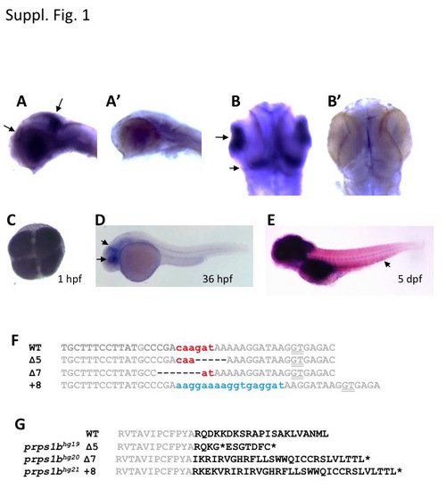
prps1 gene expression and prps1b mutations. (A-B) Flat mount images show the specific expression of prps1a in the retina and mid-brain hindbrain boundary. Wild-type embryos at 36 hpf were used for whole-mount in situ hybridization analysis with the antisense (A,B) and sense (A',B?) probes. A and A', lateral views. B and B', dorsal views. Black arrows in A and B point to the staining in the retina and brain. (C-E) prps1b expression in the wild-type embryos at the 4-cell stage (C), 36 hpf (D) and 5 dpf (E), detected by whole-mount in situ hybridization. Black arrows in D point to the enrichment in eye and tectum. Arrow in E points to the enrichment in caudal hematopoietic tissue. Images in C and D are processed for the same amount of time. Image in E is from an extended staining to show the expression in the caudal hematopoietic tissue. (F) DNA sequence alignment of the three prps1b frame-shift mutations. The wild-type (WT) DNA sequence is from Vega transcript OTTDART00000037699. ZFN target sites are shown in grey. Spacer sequence is shown in red. Deletions are shown as dashes (-). Insertions are shown in blue. Splice site GT is underlined. (G) Protein sequence alignment of the three prps1b frame-shift mutations. The WT protein sequence is from UniProt: Q08CA5_DANRE. Conserved residues are shown in grey. Premature stop codons are shown as asterisks (*). There are presumably 2, 23 and 9 extra amino acid residues introduced prior to the premature stop codon for the 5 bp deletion, 7 bp deletion, and 8 bp insertion mutations, respectively. For the 5 bp deletion mutation, there is a second premature stop codon shortly after the first one. |
| Genes: | |
|---|---|
| Fish: | |
| Anatomical Terms: | |
| Stage Range: | 4-cell to Day 5 |