
Fig. 4
- ID
- ZDB-FIG-170517-25
- Publication
- Kaslin et al., 2017 - Distinct roles of neuroepithelial-like and radial glia-like progenitor cells in cerebellar regeneration
- Other Figures
- All Figure Page
- Back to All Figure Page
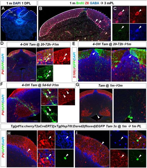
Juvenile zebrafish regenerate diverse cerebellar cell types. (A) Representative cerebellar transverse section in a juvenile 1-month-old zebrafish showing the lesion area 1 day after injury. (B,C) BrdU/ZII/GABA+ Purkinje neurons are found at the lesion site 3 months after injury (arrowheads). (D) Cerebellar transverse section showing recombined GFP/PV+ Purkinje cells (arrowheads) in Tg(ptf1a:cherryT2aCreERT2)1; Tg(hsp70l:DsRed2(floxed)EGFP) fish 1 month after recombination. (E) Cerebellar transverse section showing unilateral GFP labelling of neuroepithelial-like stem cells (arrowhead) and granule cells. (F) GFP-labelled radial glia-like cells and a clone of PV? interneurons (arrowheads) recombined between 5 and 6?days postfertilisation. (G) Quiescent GFP-labelled radial glia-like cells (arrowhead) recombined at 1 month and analysed 3 months later. (H) Recombined GFP/PV+ Purkinje cell (arrowhead) 1 month after tamoxifen treatment and injury of a juvenile zebrafish. (I) A recombined GFP+ and PV? stellate cell (arrowhead) 1 month after tamoxifen treatment and injury. Scale bars: 75 ?m (D); 35 ?m (E); 50 ?m (F); 100 ?m (G); 50 ?m (H,I). |