|
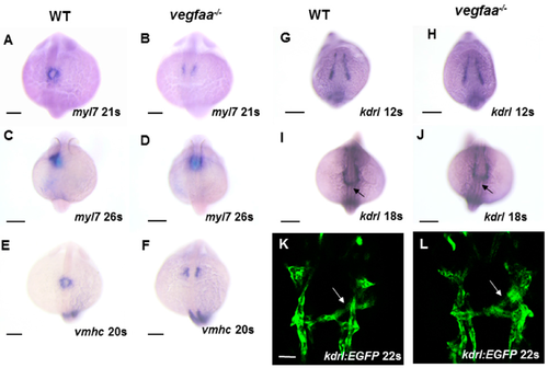
vegfaa mutants display defects in cardiomyocytes fusion. (A?D) In situ hybridization depicts myl7 expression at 21- and 26-somite stage in wild-type embryos (A,C) and vegfaa mutant embryos (B,D). At 21-somite stage, the wild-type cardiomyocytes formed a ring (A), whereas few cardiomyocytes are formed in the vegfaa mutants (B). At 26-somite stage, the wild-type primitive heart tube begins left looping (C) while vegfaa mutant embryos are still cardiac cone (D); (E,F) In situ hybridization depicts ventricular myosin heavy chain (vmhc) expression at 20-somite stage in wild-type embryos (E) and vegfaa mutant embryos (F); (G?J) Dorsal views depict unaffected endocardial expression pattern of kdrl in vegfaa mutants. Black arrows indicate midline migration of endocardial cells; Scale bar: 200 µm (A?J); (K,L) Dorsal views depict endocardial expression of Tg(kdrl:EGFP) at the 22-somite stage. White arrows indicate leftward movement. Scale bar: 50 µm.
|