
FIGURE
Fig. S2
- ID
- ZDB-FIG-170303-20
- Publication
- Kuri et al., 2017 - A high-sensitivity, bi-directional reporter to monitor NF-κB activity in cell culture and zebrafish in real-time
- Other Figures
- All Figure Page
- Back to All Figure Page
Fig. S2
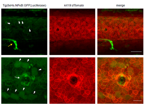
NF-κB activity in the skin of zebrafish embryos at 4dpf. Trunk region of a Tg(8xHs.NFκB:GFP,Luciferase [green]; krt19:dTomato [red]) embryo at 4dpf illustrating the sporadic NF-κB activity in the basal keratinocytes (arrowheads). Note krt19 is mainly expressed in the basal keratinocyte layer (Fischer et al., 2014). Yellow arrow indicates GFP expression in the proctodeum. White arrows indicate GFP expression in cells surrounding the lateral line. Anterior is to the left. Scale bars: 100μm (top panel) and 30μm (bottom panel). |
Expression Data
Expression Detail
Antibody Labeling
Phenotype Data
Phenotype Detail
Acknowledgments
This image is the copyrighted work of the attributed author or publisher, and
ZFIN has permission only to display this image to its users.
Additional permissions should be obtained from the applicable author or publisher of the image.
Full text @ J. Cell Sci.