|
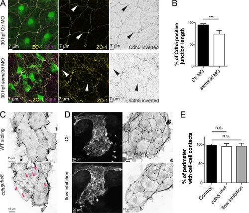
Loss of cell–cell contacts in the CCV leading edge did not depend on flow or Cdh5. (A) Antibody staining for ZO-1 and Cdh5 showed aberrant junction distribution and impaired cell shape upon sema3d loss. Arrowheads indicate gaps in Cdh5 protein distribution. (B) The length of Cdh5-positive cell–cell connections is reduced in sema3d morphants (n = 4). (C) Cell–cell connections in the CCV leading edge of cdh5ubs8 mutant embryos are present, but the ECs exhibit holes inside the cells (pink arrowheads). (D) Flow inhibition with nifedipine and tricaine did not lead to lesions or impaired Actin cable alignment in the CCV leading edge. Parallel-arranged Actin cables are indicated by open arrowheads. (E) Quantification of cell–cell-contact length of CCV leading edge cells (n = 16). ***, P < 0.001; n.s., not significant. Error bars indicate SD.
|