
Fig. 5
- ID
- ZDB-FIG-160927-11
- Publication
- Saraceni et al., 2016 - Establishment of Infection Models in Zebrafish Larvae (Danio rerio) to Study the Pathogenesis of Aeromonas hydrophila
- Other Figures
- All Figure Page
- Back to All Figure Page
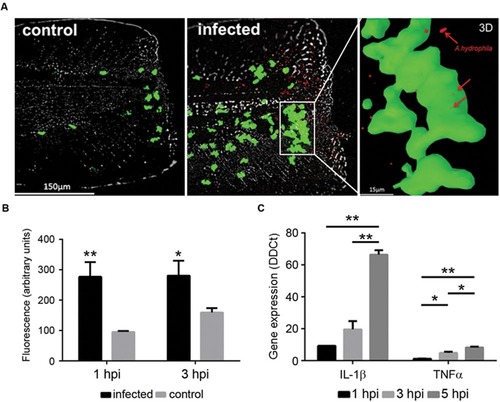
(A) In vivo imaging of neutrophils from Tg(mpx:GFP+/+) larvae after injury and bath infection with A. hydrophila (108 CFU/mL). Recruitment of neutrophils to the site of the injury 1 h after the bacterial infection. Control animals were injured but not infected. 3D reconstruction of a selected site in the infected sample showing the positions of the bacteria (red) inside the neutrophils (green). (B) Relative quantification of neutrophils in the site of injury. Results are representative of three independent experiments (n = 10-15 larvae each; ANOVA; *P < 0.05, **P < 0.01). (C) Expression of IL-1β and TNFα in injured larvae infected by bath immersion (108 CFU/mL) at 1, 3, and 5 hpi. Results of three independent experimental infections (n = 30) expressed as the mean � standard error (ANOVA and Tukey HSD test; *P < 0.05, **P < 0.01). |
| Gene: | |
|---|---|
| Fish: | |
| Condition: | |
| Anatomical Term: | |
| Stage: | Day 4 |