Fig. 3
- ID
- ZDB-FIG-160926-10
- Publication
- Thambyrajah et al., 2016 - A gene trap transposon eliminates haematopoietic expression of zebrafish Gfi1aa, but does not interfere with haematopoiesis
- Other Figures
- All Figure Page
- Back to All Figure Page
|
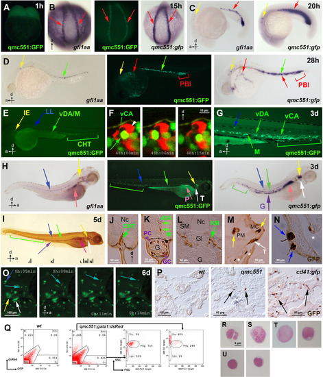
qmc551:GFP expression recapitulates gfi1aa expression and marks haematopoietic stem and progenitor cells throughout ontogeny. Views of embryos are posterior in (B) and lateral in (A,C-E,G-I,O). Anteroposterior and dorsoventral axes are indicated. (A-H) Images of live qmc551 embryos and of fixed non-transgenic and qmc551 transgenic embryos after WISH with probes against endogenous gfi1aa and gfp mRNA. (F) Confocal timelapse images (2.0 Ám thick optical slice) through the CHT of a 48 hpf qmc551;flk1:tdTom embryo. (I) Images of a qmc551 transgenic embryo immunostained for GFP expression using diaminobenzidine. (J-N). Transverse 10 Ám sections of the same embryo after plastic-embedding. The positions along the anteroposterior axis are indicated in (I). (O) Timelapse microscopy of the head region. (P) GFP immunohistochemistry and diaminobenzidine staining on 10 Ám sections of adult kidneys isolated from wt, qmc551 and cd41:gfp fish. (Q) Flow cytometric analysis of KM cells of wt and qmc551;gata1:dsRed double transgenic adults. GFP/dsRed fluorescence and forward/side scatter were analyzed. Excitation and detection wavelengths are indicated in nm. Cell populations were gated according to Traver et al. (2003). (R-U) Cytocentrifugation and Giemsa staining of the qmc551:GFP+ KM cells identified neutrophils (R), macrophages (S), progenitors (T) and lymphocytes (U). Annotations: caudal haematopoietic tissue (CHT), glomerulus (Gl), goblet cell (GC), gut (G), inner ear (IE), lateral line organ (LL), medial crista (MC), mesenchyme (M), notochord (Nc), pancreas (P), posterior blood island (PBI), putative Paneth cell (PC), pharyngeal sensory cells (asterisks), posterior macula (PM), swim bladder (SB), somitic muscle (SM), thymus (T) and ventral wall of the DA (vDA) and of the caudal artery (vCA). HSPCs, pRBCs, thymocytes and wandering leukocytes are labeled with green, red, white and turquoise arrows, respectively. |
Reprinted from Developmental Biology, 417(1), Thambyrajah, R., Ucanok, D., Jalali, M., Hough, Y., Wilkinson, R.N., McMahon, K., Moore, C., Gering, M., A gene trap transposon eliminates haematopoietic expression of zebrafish Gfi1aa, but does not interfere with haematopoiesis, 25-39, Copyright (2016) with permission from Elsevier. Full text @ Dev. Biol.

