
Fig. S9
- ID
- ZDB-FIG-160706-23
- Publication
- Raman et al., 2016 - aPKC regulates apical localization of Lgl to restrict elongation of microridges in developing zebrafish epidermis
- Other Figures
- All Figure Page
- Back to All Figure Page
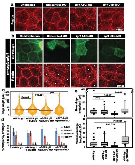
Assessment of the specificity of lgl1 morpholinos used in this study. Confocal images of microridge F-actin stained by phalloidin in un-injected embryos and embryos injected with standard control morpholino (Std control MO), lgl1ATG morpholino (lgl1ATG-MO) and lgl1UTR morpholino (lgl1UTR-MO) at 48hpf (a). Analysis of ridge lengths by F-actin staining in peridermal clones expressing eGFP-mLgl1 in embryos co-injected with the mentioned morpholino (or no morpholino) and a vector driving expression of egfp-mlgl1 under CMV promoter (b). Quantification of ridge lengths in the clones and visualisation of the length distributions and the respective medians in various genetic conditions by bean plot (c). The percentage frequency distribution of ridges in short (0-5 �m), intermediate (5-20 �m), long (20-100 �m) and very long (>100 �m) categories for the given genetic conditions (d). Estimation of cell wise means and variances of ridge lengths for each genetic condition and their representation by box-whisker plot (e). In (c) and (e), the horizontal black lines and associated probability values (P) represent the comparison by Dunn?s multiple comparisons. P<0.05 is considered statistically significant. In (d) Standard deviation is shown by error bars. Scale bars in (a,b) are equivalent to 10�m. The asterisks in (b) depict the cells that do not express eGFP-mLgl1. Abbreviation: Std: standard; MO: morphant/morpholino, mLgl1: mouse Lgl1 . |