
Fig. 2
- ID
- ZDB-FIG-160630-25
- Publication
- Lovely et al., 2016 - Bmp signaling mediates endoderm pouch morphogenesis by regulating Fgf signaling in zebrafish
- Other Figures
- All Figure Page
- Back to All Figure Page
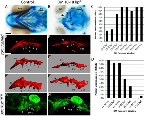
Blocking Bmp signaling early disrupts endoderm morphology and craniofacial development. (A,B) Whole-mount images of viscerocranium at 5 days post-fertilization (dpf). Cartilage is blue and bone is red. Arrowheads point to missing cartilage elements and asterisks label the ceratobranchial cartilages. (A) No craniofacial defects are present following DMSO treatment. (B) Dorsomorphin (DM) treatment from 10-18hpf causes severe craniofacial defects. Ventral views, anterior to the left. Cartilages: Mc, Meckel′s; Ch, ceratohyal; Cb′s, ceratobranchial; Pq, palatoquadrate; HM, hyomandibular. (C,D) The percentage of viscerocranial defects in embryos treated with Dorsomorphin over multiple time windows. n values for all time points are listed in Table 1. (E,F) Confocal images of control and Dorsomorphin-treated sox17:DsRed embryos. (E′-F′′) Imaris surface renderings of E,F. E′′,F′′ are rotated 45� into the plane of view. (G,H) Confocal images of control or Dorsomorphin-treated sox10:mRFP embryos. Compared with controls (E-E′′,G), Dorsomorphin-treated embryos (F-F′′,H) had severe defects to both endoderm and CNCC morphology. Lateral views (except for E′′,F′′, see above), anterior to the left. P, pouch; PA, pharyngeal arch. Scale bars: 100�m in A; 50�m in E,G. |
| Fish: | |
|---|---|
| Condition: | |
| Observed In: | |
| Stage Range: | Prim-25 to Day 5 |