
Fig. 4
- ID
- ZDB-FIG-160512-29
- Publication
- Stil et al., 2016 - Neuronal labeling patterns in the spinal cord of adult transgenic Zebrafish
- Other Figures
- All Figure Page
- Back to All Figure Page
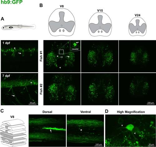
Labeling pattern of GFP in the spinal cord of adult hb9:GFP transgenic fish. In hb9:GFP larvae, GFP is driven in motoneurons and some interneurons (asterisk, A), and ventral roots (arrow, A). In adults, GFP positive fibers lie both in dorsal and ventral horns (arrow, B), and in the white matter, surrounding dlf and vlf. Some ventral commissural tracts are labeled (arrowhead, B). Cell bodies are located in the medial and the lateral parts of the ventral horn (asterisks, B, C). In the caudal part of V8, there is a strong staining above the central canal (arrows; B, inset; C). At higher magnification, GFP positive neurons show two processes (arrowheads, D). |