
Fig. 3
- ID
- ZDB-FIG-160512-28
- Publication
- Stil et al., 2016 - Neuronal labeling patterns in the spinal cord of adult transgenic Zebrafish
- Other Figures
- All Figure Page
- Back to All Figure Page
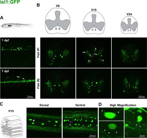
Labeling pattern of GFP in the spinal cord of adult isl1:GFP transgenic fish. In isl1:GFP larvae, transgene expression is mostly seen in secondary motoneurons (asterisk) and ventral roots (arrow, A). Transversal (B) and coronal (C) sections of adult spinal cord show large GFP-positive cell bodies in the dorsal part (white asterisks), and smaller cells more ventrally (magenta asterisks). Processes (arrow, B) extend in the upper region of ventral horns. The start of ventral roots (arrowheads, B) is stained. No signal is detected in the dorsal part. Higher magnification highlights large and smaller cells with two or more processes (D). |