
Fig. 6
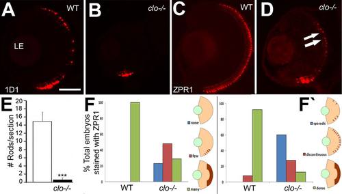
Reduced and mispatterned differentiation of photoreceptors in cloche mutant embryos. A,B: Immunofluorescence images of 72 hpf retinas of wild-type (A) and clo-/- (B) embryos stained with the 1D1 antibody, detecting rod photoreceptors. C,D: Immunofluorescence images of 72 hpf retinas of wild-type (D) and clo-/- (E) embryos stained with the ZPR1 antibody, detecting cone photoreceptors. Arrows in D show developing cones flanking a region of the ONL that is not ZPR1+. E: Numbers of 1D1+ cells are significantly (***P < 0.001) reduced in clo-/- retinas. F,F′: The extent of ZPR1 labeling (F) and the distribution of ZPR1+ cells (F2) is also significantly different in clo-/- retinas as compared to wild-type (P < < 0.001). LE, lens. Scale bar = 50 �m in A (applies to all images). |