
Fig. 5
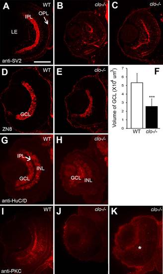
Abnormal differentiation of specific inner retinal neurons in clo mutant embryos. A?C: Immunofluorescence images of 72 hpf retinas of wild-type (A) and clo-/- (B,C) embryos stained for synaptic vesicle 2 (SV2). D,E: Immunofluorescence images of 72 hpf retinas of wild-type (D) and clo-/- (E) embryos stained with the ZN8 antibody, detecting retinal ganglion cells. F: Volume of GCL is significantly (***P < 0.001) reduced in clo-/- retinas. G,H: Immunofluorescence images of 72 hpf retinas of wild-type (G) and clo-/- (H) embryos stained with an antibody detecting HuC/D, present in ganglion cells and amacrine cells. I?K: Immunofluorescence images of 72 hpf retinas of wild-type (I) and clo-/- (J,K) embryos stained with an antibody detecting PKCα, specific to bipolar cells. * in K indicates diffuse staining of material lacking typical bipolar cell morphology. Scale bar = 50 �m in A (applies to all images). LE, lens; IPL, inner plexiform layer; OPL, outer plexiform layer; GCL, ganglion cell layer; INL, inner nuclear layer. |