
Fig. S6
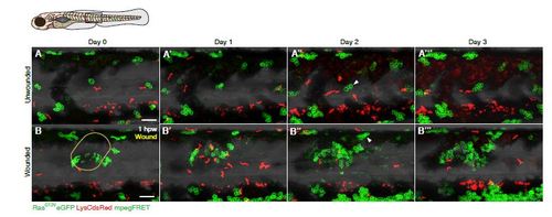
Timecourse of clonal growth in the presence versus absence of a wound. A-B A series of time-lapse images showing clonal growth in a wounded RasG12VeGFP LysCdsRed mpegFRET larva and unwounded control over a period of four consecutive days. (A) Control, unwounded larva, versus (B) Larva wounded at 4dpf. Arrowhead in (B′′) highlights a clone close to the wound, which has increased in size from two to seven cells over a 24 hour period. Over the same period, most clones in the control larva grow more slowly (arrowhead in A′′). The approximate domain for imaging is indicated by a box in the schematic. Scale bars represent 100µm. |