
FIGURE
Fig. S1
Fig. S1
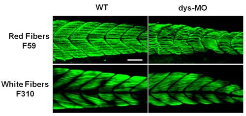
Immunostaining distinguished the red and white fibers of skeletal muscles. Red fibers were labeled by F59 antibody for slow myosin (n=20) and white fibers were labeled by F310 antibody for fast myosin (n=18). Distinctive mitochondrial morphologies were found in these fiber types at 2 dpf as shown in Figure B and S1 Movie. Note abnormalities of these fibers in dys-MO-injected morphants. Scale bar, 50 �m. |
Expression Data
Expression Detail
Antibody Labeling
Phenotype Data
Phenotype Detail
Acknowledgments
This image is the copyrighted work of the attributed author or publisher, and
ZFIN has permission only to display this image to its users.
Additional permissions should be obtained from the applicable author or publisher of the image.
Full text @ PLoS One