
Fig. 3
- ID
- ZDB-FIG-150609-17
- Publication
- B³hrdel et al., 2015 - In vivo characterization of human myofibrillar myopathy genes in zebrafish
- Other Figures
- All Figure Page
- Back to All Figure Page
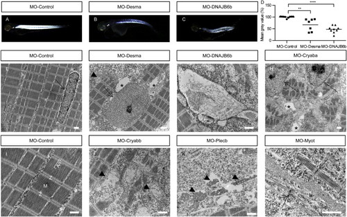
Disturbed myofibrillar structure in MFM gene deficient embryos (A?C) Lateral views of a control zebrafish (A) at 72 hpf showing bright homogenous birefringence compared to reduced birefringence in Desmin-a (B) and DNAJB6-b (C) morphants. (D) Quantification of birefringence in Desmin-a and DNAJB6-b morphants. (E?L) Electron microscopy of MFM gene deficient and control embryos at 72 hpf. (E) Nuclei (dotted arrows) in the skeletal muscle are elongated in controls; (F,H) rounded nuclei in myopathic muscle. (I) Control injected embryos develop highly ordered myofibrils with ordered sarcomeric units, Z-disks, M-Bands and A/I regions. (F,H) Vacuolization in MO-Desma and MO-crayaba injected embryos (asterisk). (G) Autophagic pathology in Dnajb6-a morphants. (F,J,K) Mitochondrial alteration in Desmin-a, Plectin-b and αB-crystallin-a morphants (arrowheads). (L) Stress fiber-like structures in Myotilin morphants (arrows). Scale bars 1 Ąm. |
| Fish: | |
|---|---|
| Knockdown Reagents: | |
| Observed In: | |
| Stage: | Protruding-mouth |