
Fig. 3
- ID
- ZDB-FIG-150505-6
- Publication
- Lin et al., 2015 - Angiopoietin-like proteins stimulate HSPC development through interaction with Notch receptor signaling
- Other Figures
- All Figure Page
- Back to All Figure Page
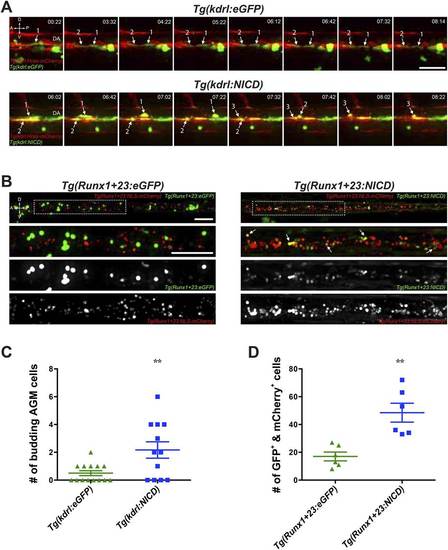
Notch cell autonomously increase definitive hematopoiesis. (A) Time-lapse sequence (hours:minutes post-28hpf) of HSPCs emerging from the ventral wall of the DA. Hemogenic endothelial cells (red) that have incorporated the injected transgene (green) are marked with numbers and white arrows. Each injected embryo was scored for 24 hr and tabulated in (C). Scale bar: 25 �m. (B) Still images of the CHT from 72hpf embryos. Runx1-positive HSPCs (red) that have incorporated the injected transgene (green) were scored for double positivity (yellow, examples marked by white arrows) and tabulated in (D). Boxed areas in the top panels are magnified and split into double or single fluorescent panels below. Scale bars: 50 �m. A: Anterior, P: Posterior, D: Dorsal; V: Ventral. Error bars denote S.E.M., **p < 0.01, compared to eGFP injected controls, Student′s t test. |