
FIGURE
Fig. 5
- ID
- ZDB-FIG-150423-27
- Publication
- Williams et al., 2015 - MASH1/Ascl1a Leads to GAP43 Expression and Axon Regeneration in the Adult CNS
- Other Figures
- All Figure Page
- Back to All Figure Page
Fig. 5
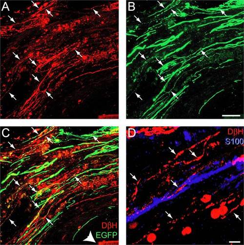
Rats treated with MASH1 exhibit EGFP expression in noradrenergic axons that regenerated into a SC bridge. A-C, Expression of DβH (red) and EGFP (green) was sometimes co-localized (arrows) in axons that regenerated past the rostral spinal cord/SC bridge interface. Arrowhead indicates the polymer channel (scale bar = 20 �m). D, DβH-positive axons (red, arrows) regenerated in close proximity to S100-positive SCs (blue; scale bar = 5 �m). |
Expression Data
Expression Detail
Antibody Labeling
Phenotype Data
Phenotype Detail
Acknowledgments
This image is the copyrighted work of the attributed author or publisher, and
ZFIN has permission only to display this image to its users.
Additional permissions should be obtained from the applicable author or publisher of the image.
Full text @ PLoS One