
Fig. S5
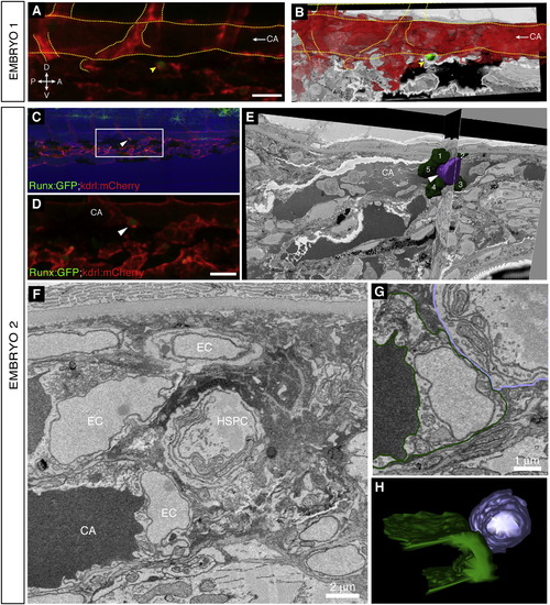
High-Resolution Electron Microscopy of Endogenous HSPC in the Perivascular Niche, Related to Figure 6 (A and B) Embryo 1 (same embryo as shown in Figure 6). Comparison of 3D projections from (A) confocal z-stack data and (B) serial EM scans to confirm the position of the lodged HSPC. (A) 3D rendered projection of z-stack (Imaris software) showing one lodged Runx:GFP+ HSPC (green; arrowhead) and kdrl:mCherry+ ECs (red). Scale bar: 25 �m. (B) Vessel lumen manually outlined, surface rendered, and shown in red (Imaris software). 3D projection overlaid on a single EM section. Rotation performed to match orientation of image in (A). Orientation: D (dorsal), V (ventral), A (anterior), P (posterior). CA (caudal artery). Circulation is toward the posterior (left; arrow). Major vessels outlined. (C?H) Embryo 2. A second independent example of serial block face EM sections on a lodged HSPC after time-lapse. (C) Last frame of CHT time-lapse (60 hpf). Arrowhead marks HSPC lodged > 6 hr. Runx:GFP green; kdrl:mCherry red; bright field blue. Anterior left, posterior right, dorsal top, ventral bottom. (D) Detail of region in (C) marked by box. The brightness of the GFP channel was adjusted independently of the mCherry channel to clearly show the position of the Runx:GFP+ HSPC. Scale bar: 25 �m. (E) Single section and orthogonal slice from serial block face EM scans. Lodged HSPC is purple (arrowhead), surrounding EC nuclei are green and numbered. Part of the HSPC is missing because it was not captured in serial EM sections. (F) High resolution EM section of HSPC in perivascular niche with cell nuclei labeled. (G) EM section with cell membranes outlined to show contact between EC (dark green) and HSPC (purple). (H) 3D modeling shows one side of the HSPC is in contact with the EC. For animated 3D reconstructions see Movie S7. |
Reprinted from Cell, 160, Tamplin, O.J., Durand, E.M., Carr, L.A., Childs, S.J., Hagedorn, E.J., Li, P., Yzaguirre, A.D., Speck, N.A., Zon, L.I., Hematopoietic Stem Cell Arrival Triggers Dynamic Remodeling of the Perivascular Niche, 241-252, Copyright (2015) with permission from Elsevier. Full text @ Cell