
Fig. 2
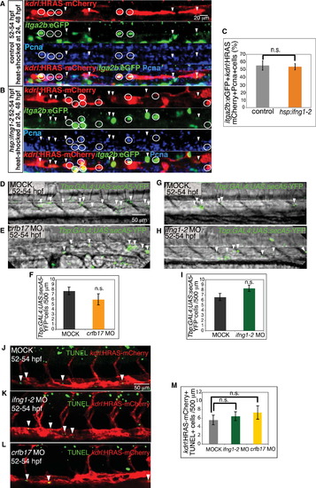
Cell Proliferation and Survival in the Axial Vessels Do Not Appear to Be Affected by Gain or Loss of Function of Ifng1-2 Signaling (A?C) ifng1-2 overexpression increases HSC number without affecting cell division. HSCs (itga2b:EGFP+kdrl:HRAS-mCherry+, green+red+) that exit the G0 phase, labeled by PCNA immunostaining (blue), in control embryos not harboring the Tg(hsp:ifng1-2-V5) transgene (A), and Tg(hsp:ifng1-2-V5) embryos (B) are indicated by white circles. PCNA HSCs are marked with white arrowheads. (C) Percentage of PCNA+HSCs per total HSCs in the dorsal aorta. Values represent means � SEM, n = 9 embryos per group, n.s., not significant (p > 0.05). (D?M) Ifng1-2 and Crfb17 knockdown has no effect on apoptosis. Apoptotic cells (green, white arrowheads) visualized by Tg(Tbp:GAL4;UAS:secA5-YFP) expression (D?I) and TUNEL assay (J?M) in the axial vessels of MOCK-injected (D, G, and J), crfb17 MO-injected (E and L), and Ifng1-2 MO-injected (H and K) embryos at 52?54 hpf. Numbers of Tbp:GAL4;UAS:secA5-YFP+ cells (F and I) and TUNEL+kdrl:HRAS-mCherry+ cells per 500 �m dorsal aorta length (M) are shown as means � SEM, n = 11?25 embryos, n.s. p > 0.05. All images are lateral views, dorsal up and anterior to the left. |
| Genes: | |
|---|---|
| Antibody: | |
| Fish: | |
| Condition: | |
| Knockdown Reagents: | |
| Anatomical Terms: | |
| Stage: | Long-pec |
Reprinted from Developmental Cell, 31, Sawamiphak, S., Kontarakis, Z., Stainier, D.Y., Interferon Gamma Signaling Positively Regulates Hematopoietic Stem Cell Emergence, 640-653, Copyright (2014) with permission from Elsevier. Full text @ Dev. Cell