Fig. 1
- ID
- ZDB-FIG-150108-1
- Publication
- Huang et al., 2013 - Sequential effects of spadetail, one-eyed pinhead and no tail on midline convergence of nephric primordia during zebrafish embryogenesis
- Other Figures
- All Figure Page
- Back to All Figure Page
|
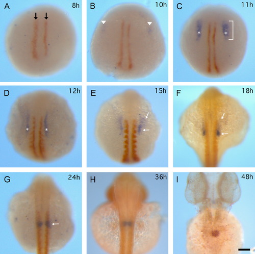
Morphogenetic changes of PGP during 8-48 hpf development. Dynamic wt1a expression (blue) in relation to somite morphogenesis (myoD expression, orange-brown) at 8, 10, 11, 12, 15, 18, 24 hpf is shown in (A)-(G). (A) MyoD expression labels adaxial cells at 8 hpf (black arrow). (B) Initial wt1a expression (arrowhead) locates bilaterally at a distance from the adaxial cells. ((C) and (D)) Wt1a expression pattern expands along the (A)-(P) dimension (bracket). Wt1a expression crosses on left-right axis at third somite were the measure points for inter distance of PGP (asterisks). ((E) and (F)) White arrows indicate faint (anterior side) and dense (posterior side) signal in wt1a-positive cells. (G) The faint signal is almost unseen and wt1a-positive cells become compact round shape (white arrow). (H) The bilateral PGP (blue) adjacent to each other at 36 hpf, myoD and mibp2 were stained orange-brown. (I) A single fuse PGP (orange-brown) appears in midline at 48 hpf. All images are dorsal view and at the same magnification. The scale bar in H indicates 100 Ám. |
| Genes: | |
|---|---|
| Fish: | |
| Anatomical Terms: | |
| Stage Range: | 75%-epiboly to Long-pec |
Reprinted from Developmental Biology, 384(2), Huang, C.J., Wilson, V., Pennings, S., MacRae, C.A., and Mullins, J., Sequential effects of spadetail, one-eyed pinhead and no tail on midline convergence of nephric primordia during zebrafish embryogenesis, 290-300, Copyright (2013) with permission from Elsevier. Full text @ Dev. Biol.

