
Fig. 2
- ID
- ZDB-FIG-140819-2
- Publication
- Trevarrow et al., 1990 - Organization of hindbrain segments in the zebrafish embryo
- Other Figures
- All Figure Page
- Back to All Figure Page
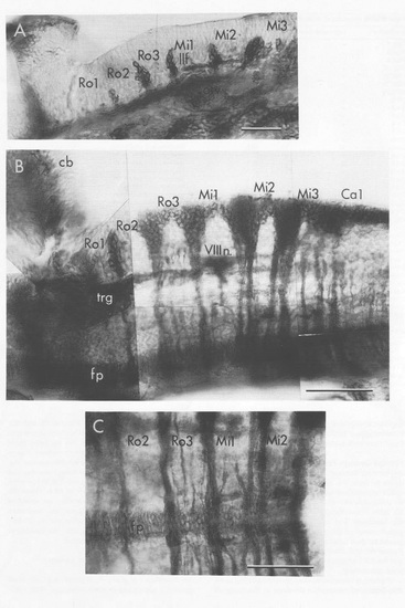
The Zn-5 Antibody Labels Clusters of Neurons and Their Commissural Axons That Develop Near the Segment Borders (A) Sagittal section at 29 h. The clusters are located dorso-lateral to the lateral longitudinal fasciculus (llf, a predominantly sensory pathway that includes descending fibers of the trigeminal nerve; see P.Z. Myers, W.K. Metcalfe, and C.B. Kimmel, submitted), revealing their location in the alar plate. The developing pharyngeal arches ventral to the hindbrain and the otic vesicle (otv) are also labeled. (B and C) Views of the hindbrain in 48h embryos dissected after fixation. (B) Dorsolateral oblique view. The labeled cell clusters lie in the border regions dorsal (aand lateral) to the eighth nerve fibers (VIIIn.), which enter the brain in the Mi1 segment. Note that the neuronal clusters in the Mi1-2, Mi2-3, and Mi3-Ca1 border regions project two large bundles of commissural axons, as contrasted with the clusters located more rostrally, which project only a single large bundle. The floor plate cells (fp) at the ventral midline form a continuous, nonsegmented row in the hindbrain (and also in the midbrain; data not shown). cb: cerebellum, trg:trigeminal ganglion. (C) Dorsal view of the midline region. The double commissures in (B) and (C) are in the same locations. In addtion, one or two labeled commissures extend across the centers of some segments (e.g. Ro3). Scale bars: 50 μm. |
| Antibody: | |
|---|---|
| Fish: | |
| Anatomical Terms: | |
| Stage Range: | Prim-5 to Long-pec |