
Fig. 1
- ID
- ZDB-FIG-140819-1
- Publication
- Trevarrow et al., 1990 - Organization of hindbrain segments in the zebrafish embryo
- Other Figures
- All Figure Page
- Back to All Figure Page
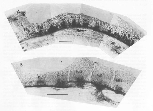
Antibodies Reveal the Segmental Organization of the Embryonic Zebrafish Hindbrain (A) Labeling by the zn-1 antibody, which recognizes many or all neurons (Table 1). A cluster of labeled cells occupies the center of each neuromere, and much smaller groups of positive cells are beginning to develop in the border regions between some of the neuromeres. The hindbrain-midbrain (cerebellar region: cb) boundary is shown to the left. The fourth ventricle is dorsal to most of the hindbrain tissue. (B) Labeling by the zn-12 antibody, which at early stages recognizes a subset of neurons present in the segment centers. Sagittal sections at 24 h are shown rostral to the left and dorsal up. All subsequent figures showing sagittal views are oriented the same way. Here and throughout the paper, naming of the hindbrain segments follows previous conventions (Metcalfe et al., 1986; Hanneman et al. 1988); there are three rostral segments, Ro1-Ro3, three middle ones, Mi1-Mi3, and three caudal divisions, Ca1-Ca3. Scale bars: 50 μm. |
| Antibodies: | |
|---|---|
| Fish: | |
| Anatomical Term: | |
| Stage: | Prim-5 |