
Fig. 5
- ID
- ZDB-FIG-140523-41
- Publication
- Soza-Ried et al., 2014 - Pulses of Notch activation synchronise oscillating somite cells and entrain the zebrafish segmentation clock
- Other Figures
- All Figure Page
- Back to All Figure Page
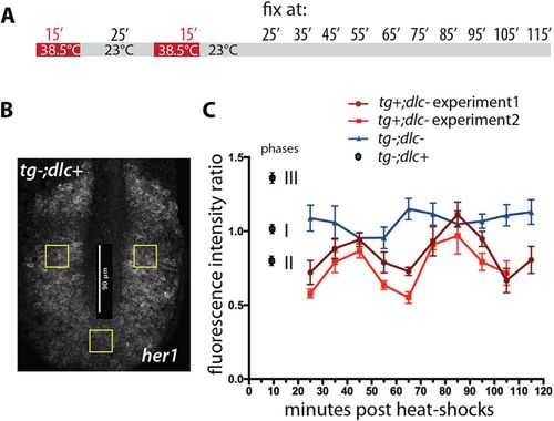
Rescued somite boundary formation correlates with rescued coordination of her1 oscillation. Mutant tg+;dlc embryos were taken at the 12-somite stage, given two standard 38.5�C heat-shocks separated by 25min at 23�C, and then maintained at 23�C for varying lengths of time before fixation. (A) Scheme of the treatment. (B) Wild-type embryo stained by FISH with her1 riboprobe, indicating the regions chosen for measurement (30�30�m2 yellow boxes) for all specimens in this experiment. Scale bar (vertical): 90μm. Fluorescence intensity was measured photometrically from z-stack projections. As an indicator of coordinated her1 oscillation, we took the ratio of the level of her1 in the extreme posterior box to the levels in the two symmetrically placed more anterior (mid-PSM) boxes. (C) Graph of this ratio as a function of time after the end of the second heat-shock. Two repetitions of the whole experiment are shown (brown and red lines), along with corresponding measurements for control tgdlc embryos (blue line). Each data point is an average over at least three, and on average 5.4, specimens; error bars show s.e.m. Both repetitions of the heat-shock experiment show similar clear oscillations, with period of <40min, as expected for the segmentation clock at 23�C (Schroter et al., 2008). By contrast, the control series of deltaC/ embryos without heat-shock shows only small fluctuations. The measurements for control tgdlc+ embryos are at the left-hand side of the plot, and assigned to cycling phase I, II or III [nomenclature of Pourquie and Tam (Pourquie and am, 2001)]. |
| Gene: | |
|---|---|
| Fish: | |
| Anatomical Term: | |
| Stage Range: | 10-13 somites to 14-19 somites |