
Fig. S3
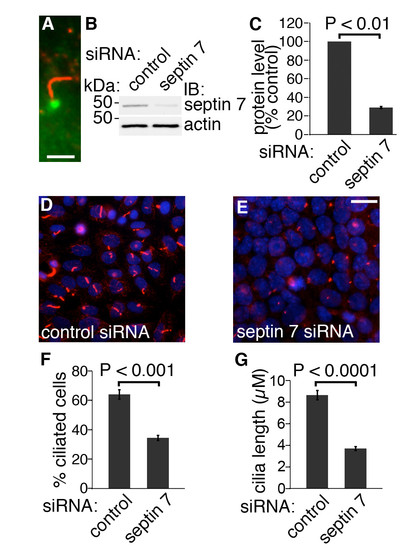
Knockdown of septin 7 decreases the length of cilia in mIMCD3 cells. (A) In mIMCD3 cells, septin 7 (green) is expressed at the base of cilia, which are stained for acetylated tubulin (red). Scale bar: 2,5 μm. (B) mIMCD3 cells were transfected with 100 nmol ON-TARGET plus SMARTpool mouse Sept7 (L-042160-01-0005) or siCONTROL Non-Targeting Pool#2 (D-001206- 14-05) siRNAs (Dharmacon, Lafayette, CO) using Lipofectamine 2000 (Invitrogen). Septin 7 siRNA leads to a 71% reduction in septin 7 expression in mIMCD3 cells. Actin was used as a loading control. (C) Quantification of protein levels of three replicate blots as in (B). (D-E) mIMCD3 cells treated with control siRNA (D) are decorated with cilia identified by staining for acetylated tubulin (red). In mIMCD3 cells transfected with septin 7 siRNA (E), the number and length of cilia are reduced. Nuclei are visualized with Hoechst stain (blue). Scale bar: 10 μm. (F) Quantification of 4000 cells indicates that 64% of cells transfected with the control siRNA had cilia whereas only 34.5% of cells transfected with septin 7 siRNA were ciliated. (G) Cilia length is reduced in septin 7 siRNA treated cells (3.69 � 0.47 μm) compared to control siRNA transfected cells (8.86 � 0.63 μm; n=200 for both). Graphs in C, F and G show the mean and error bars (STDEV) of three independent experiments, Student?s t-test. |