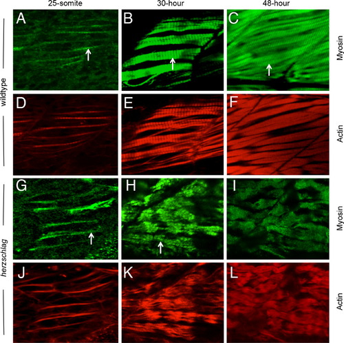
Myosin thick filament organization still occurs in the absence of the titin rod domain. Herzschlag mutants and wild-type embryos were followed through a time-course of myogenesis, and stained at 24, 30 and 48 hpf with antibodies against slow-muscle myosin (green). In wild-type embryos (A?F), the striations of organized thick filaments can be seen at all time-points throughout myofibrillogenesis (arrows in A?C). Counter-staining with fluorescently labeled phalloidin to show actin organization (red) reveals a similar pattern of thin filament organization (D?F). In hel mutant embryos, the striated pattern of myosin thick filaments was retained at 24 h (G, arrow). By 30 hpf, myofibers were clearly disorganized (H), but retained a striated pattern of thick filament myosin (arrow). This pattern was only lost completely after 48 hpf (I). Thin filament striations, as show by phalloidin counter-staining, were never visible at any timepoint in hel mutant embryos (J?L). In all experiments, a minimum of 20 embryos were examined to ensure consistency of the phenotype.
|
