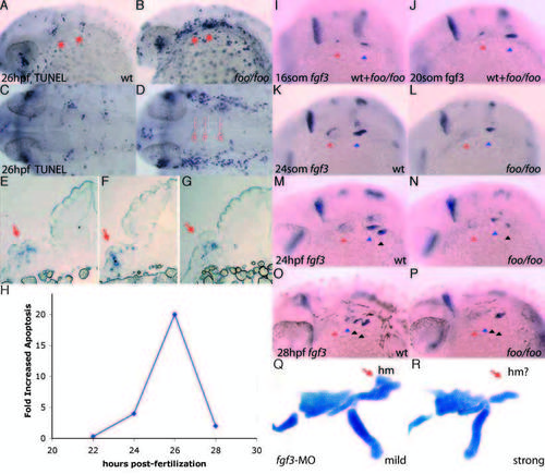
foo mutants show a loss of fgf3 expression maintenance in the pharyngeal pouches and increased apoptosis. (A,B) TUNEL assay on a wild-type (A) and foo/foo (B) embryos at 28 hpf, lateral views. Arrows indicate pp1 and pp2 with increased apoptosis. (C) Dorsal view of A; (D) dorsal view of B. Broken lines indicate approximate section locations. (E-G) Sections of TUNEL assay stained foo/foo embryos indicated in D. Arrows indicate TUNEL-positive cells. (H) Transient increase in apoptosis observed in foo/foo embryos. Quantitation was performed using NIH-Image for densitometry analysis of corresponding arch regions between mutant and wild-type embryos. Data points represent averages for at least three embryos. (I-P) fgf3 expression by in situ hybridization in foo/foo (I,J,L,N,P) and wild-type (I,J,K,M,O) siblings. (I) 16- somite stage (mutants are indistinguishable from wild type at this stage). (J) 20- somite stage (mutants can not be distinguished from wild type). (K) 24-somite stage embryo. fgf3 expression is maintained in pp1 (red arrowhead) and pp2 (blue arrowhead). (L) 24-somite stage. fgf3 expression maintenance is lost from pp1, while pp2 expression is slightly lower than in wild type (K), but clearly visible. (M) 24 hpf embryo. fgf3 expression is maintained in pp2 (blue arrowhead) and pp3 (black arrowhead). (N) 24 hpf embryo. fgf3 expression maintenance is reduced from pp2 and initiation is visible in pp3. (O) 28 hpf embryo. fgf3 expression is absent from pp1 (red arrowhead), is downregulated in pp2 (blue arrowhead), maintained in pp3 (left black arrowhead) and initiated in pp4 (right black arrowhead). (P) 28 hpf embryo. Expression is initiated normally in pp4 (second black arrowhead) but is prematurely lost from pp2 (blue arrowhead) and pp3 (left black arrowhead). (Q) Flat-mounted Alcian Blue staining of a mildly affected 4 dpf embryo injected with 5 ng of fgf3-MO (Phillips et al., 2001). Hyosymplectic (red arrow) appears relatively normal. (R) A more strongly affected 4 dpf embryo injected with 5 ng of fgf3-MO shows a reduction in the hyosymplectic (red arrow) consistent with the skeletal defects observed in foo/foo embryos.
|

