|
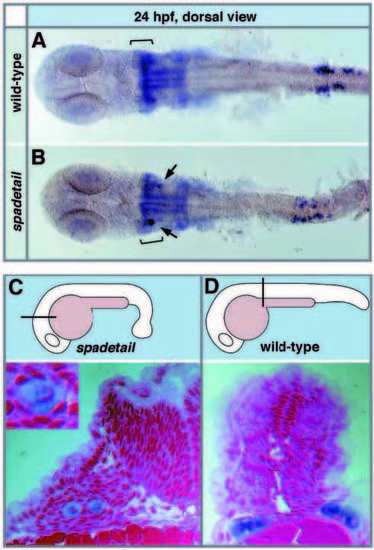
PGCs maintain their fate at ectopic locations. All embryos were stained with the vasa probe in blue. (A, B) Double staining of wild-type (A) and spadetail mutant (B) embryos with vasa and hoxa2 both in blue shows that anterior ectopic PGCs (arrows) are located at the anteroposterior level of the 2nd branchial arch (brackets) at 24 hpf. (C, D) Ectopic vasa-expressing cells maintain PGC morphology. Cross-section of a spadetail mutant (C) and a wild-type (D) embryo at the indicated levels. Note that both the ectopic vasa-positive cells in C and the PGCs located in the correct position in D are large and show a distinct nuclear shape (see insert).
|