
Fig. 5
- ID
- ZDB-FIG-140227-42
- Publication
- Macdonald et al., 1995 - Midline signalling is required for Pax gene regulation and patterning of the eyes
- Other Figures
- All Figure Page
- Back to All Figure Page
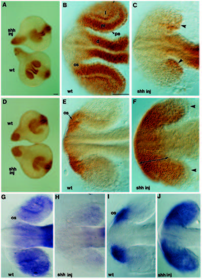
shh has opposite effects upon Pax2 and Pax6 expression in the optic primordia. Whole-mounted 16-18s embryos labelled with antibodies to Pax6 (A-C) or Pax2 (D-F), or hybridised with antisense RNA probes to pax6 (G,H) or pax2 (I,J). Rostral CNS is to the left. (A) Dorsal views of Pax6 distribution in shh-injected (upper) and wild-type (lower) embryos. (B,C) Higher magnification views showing Pax6 distribution in the forebrain and eyes of wildtype (B) and injected (C) embryos. Some anti-Pax6 antibody labelling persists in the caudal portions of the optic primordia of the injected embryo (arrowheads in C). (D) Dorsal view of Pax2 distribution in intact wild-type (upper) and injected (lower) embryos. (E,F) Higher magnification views showing Pax2 distribution in the forebrain and optic primordia of wild-type (E) and injected (F) embryos. The most caudal cells of the optic primordia of the injected embryo are not labelled with the anti-Pax2 antibody (arrowheads in F). The area of attachment of the optic primordia to the diencephalon is greatly increased in the shh-injected embryo (double headed arrow in F). (G,H) Dorsal view of pax6 expression in the eye primordia of wild-type (G) and shh-injected (H) embryos. (I,J) pax2 expression in the optic primordia of wild-type (I) and shh-injected (J) embryos. Abbreviations: d, diencephalon; l, lens; nr, neural retina; os, optic stalk; pe, pigment epithelium. Scale bars, 50 μm. |